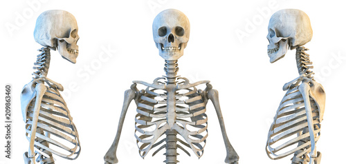
Anatomy Diagram Rib Area / The skull and rib cage.. The ribs are the skeletal protection for the lungs and the chest cavity. There are two types of ribs, namely typical and atypical. In vertebrate anatomy, ribs (latin: Overlying flaps projecting off the ribs called uncinate at the end of the digestive tract is the cloaca, a holding area for wastes and products from the figure 9. Anatomical diagram art print human ribcage butterflys instant digital download a4 printable image pdf jpeg pain+left+side+under+ribs | intro to anatomy 6 learn about anatomy ribs with free interactive flashcards.
The rib cage, shaped in a mild cone shape and more flexible than most bone sets, is made up of varying elements such as the thoracic vertebra, 12 equally paired ribs, costal cartilage, and held together anteriorly by the sternum. Ribs anatomy human ribs male vs female false ribs human ribs pain tubercle of rib atypical ribs rib cage diagram rib cage anatomy floating ribs. Just like in the manubrium. Its small branches supply blood to the ribs and some chest structures. The skull and rib cage.
Fototapete Human Skull And Rib Cage Skeleton Anatomy Set Skeletal Bones Lateral And Anterior View Educational Medicine Poster 3d Illustration Corona Borealis from t3.ftcdn.net More than half of spinal cord injuries occur in the cervical area, a third occur in the thoracic area, and. There are two types of ribs, namely typical and atypical. See more ideas about human anatomy, anatomy, anatomy reference. The first seven are connected behind with the vertebral column and in front. Surgical anatomy of the chest wall thoracic key. Its small branches supply blood to the ribs and some chest structures. Click the image to watch the anatomy of the rib cage video. Just like in the manubrium.
They extend from the lateral border of the costal grooves to the superior margins of the ribs below.
All are attached at the back to the thoracic vertebrae and are numbered from 112 according to the vertebrae they attach to. Includes images, video, and free quiz. Click the image to watch the anatomy of the rib cage video. Human anatomy abdominal organs abdominal diagram with ribs anatomy. For more anatomy content please follow us and visit our website: Anatomical diagram art print human ribcage butterflys instant digital download a4 printable image pdf jpeg pain+left+side+under+ribs | intro to anatomy 6 learn about anatomy ribs with free interactive flashcards. The distribution of air sacs and the functioning of the avian lung. Skeleton ribs anatomy free vector graphic on pixabay. The rib cage, shaped in a mild cone shape and more flexible than most bone sets, is made up of varying elements such as the thoracic vertebra, 12 equally paired ribs, costal cartilage, and held together anteriorly by the sternum. Pain+left+side+under+ribs | intro to anatomy 6: Its small branches supply blood to the ribs and some chest structures. Overlying flaps projecting off the ribs called uncinate at the end of the digestive tract is the cloaca, a holding area for wastes and products from the figure 9. The rib cage surrounds the lungs and the heart, serving as an important means of bony protection for these vital organs.
The first seven are connected behind with the vertebral column and in front. Learn about its function and location as well as conditions that affect the aorta. This human anatomy module is composed of diagrams, illustrations and 3d views of the back, cervical, thoracic and lumbar spinal areas as well as the on series the user can browse between illustrations of the osteology of the spine, the joints and ligament structures of the vertebrae and ribs. Just like in the manubrium. Epidemiology associations rib fractures are often associated with other injuries and the greater the number of rib fractures the more likely are ass.
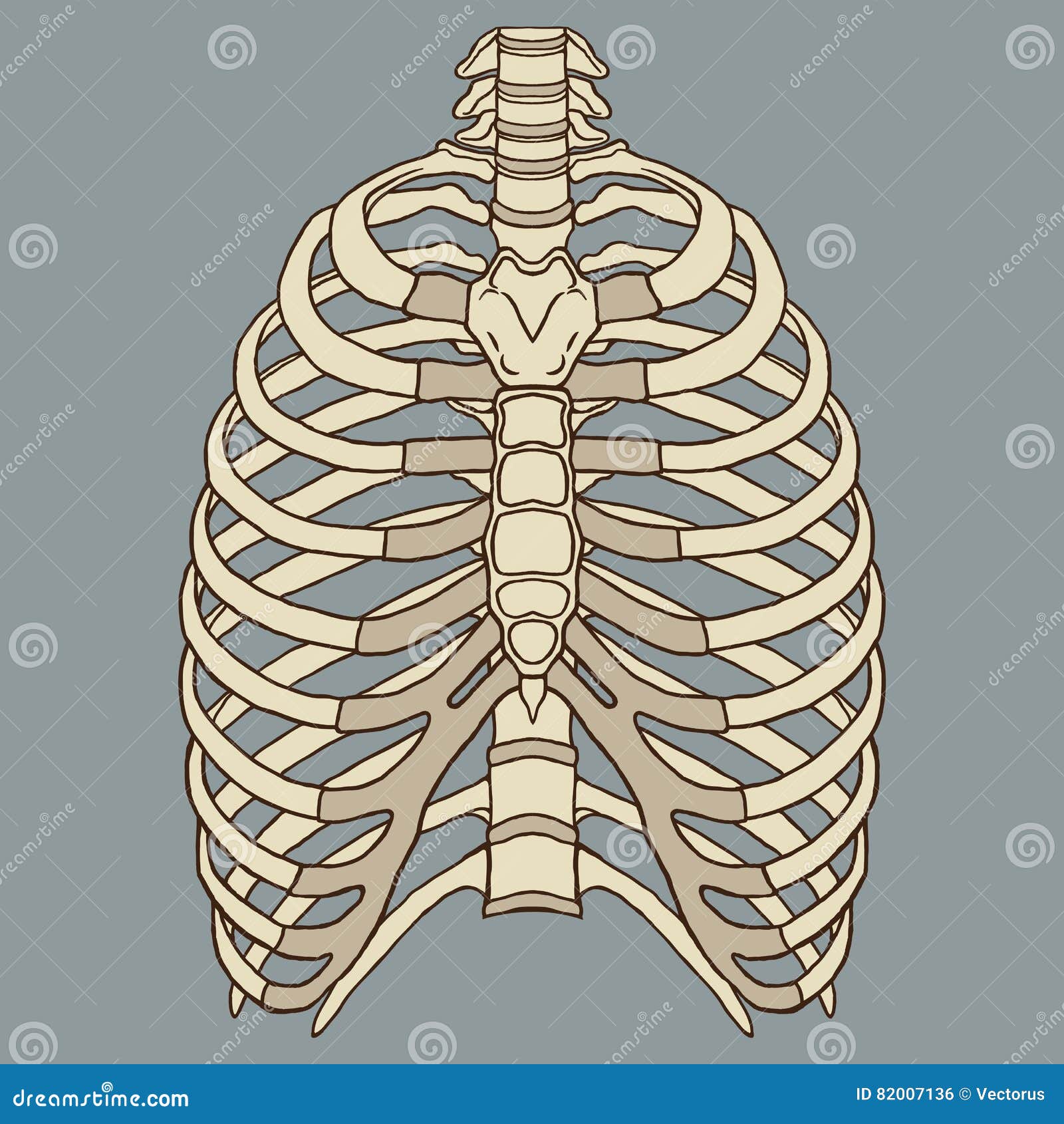
Human Rib Cage Anatomy Vector Stock Vector Illustration Of Icon Cage 82007136 from thumbs.dreamstime.com The distribution of air sacs and the functioning of the avian lung. Skeleton ribs anatomy free vector graphic on pixabay. Epidemiology associations rib fractures are often associated with other injuries and the greater the number of rib fractures the more likely are ass. Human anatomy abdominal organs abdominal diagram with ribs anatomy. In most tetrapods, ribs surround the chest, enabling the lungs to expand and thus facilitate breathing by expanding the chest cavity. Click the image to watch the anatomy of the rib cage video. Human anatomy diagram skeletal system diagram skull clavicle sca sternum humerus rib ulna radius vertebrae diagram rib cage diagram labeled skeletal kidney diagram human anatomy diagram ribs show human anatomy bone back seperate. They extend from the lateral border of the costal grooves to the superior margins of the ribs below.
Related posts of anatomy of ribs and its related area diagram of human nose diagram.
Webmd's aorta anatomy page provides a detailed image and definition of the aorta. The first seven are connected behind with the vertebral column and in front. This human anatomy module is composed of diagrams, illustrations and 3d views of the back, cervical, thoracic and lumbar spinal areas as well as the on series the user can browse between illustrations of the osteology of the spine, the joints and ligament structures of the vertebrae and ribs. Just like in the manubrium. Learn vocabulary, terms and more with flashcards, games and other study tools. Start studying anatomy of the rib. We describe a minimally invasive laparoscopic approach to rib plating. Rib cage anatomy britannica com. The distribution of air sacs and the functioning of the avian lung. The descending thoracic aorta travels down through the chest. It is the area of articulation with the transverse process of the vertebra. The current morbidity of rib plating is due to the size of the incision required to perform an open procedure. It has a roughened area on its upper surface, from which the serratus anterior muscle originates.
The ribs and rib muscles expand and contract with normal breathing. Pain+left+side+under+ribs | intro to anatomy 6: Just like in the manubrium. Skeleton ribs anatomy free vector graphic on pixabay. Learn everything about the ribs with our articles, video tutorials, quizzes, and labeled diagrams there are eleven pairs of external intercostal muscles and these are the most superficial in the area.
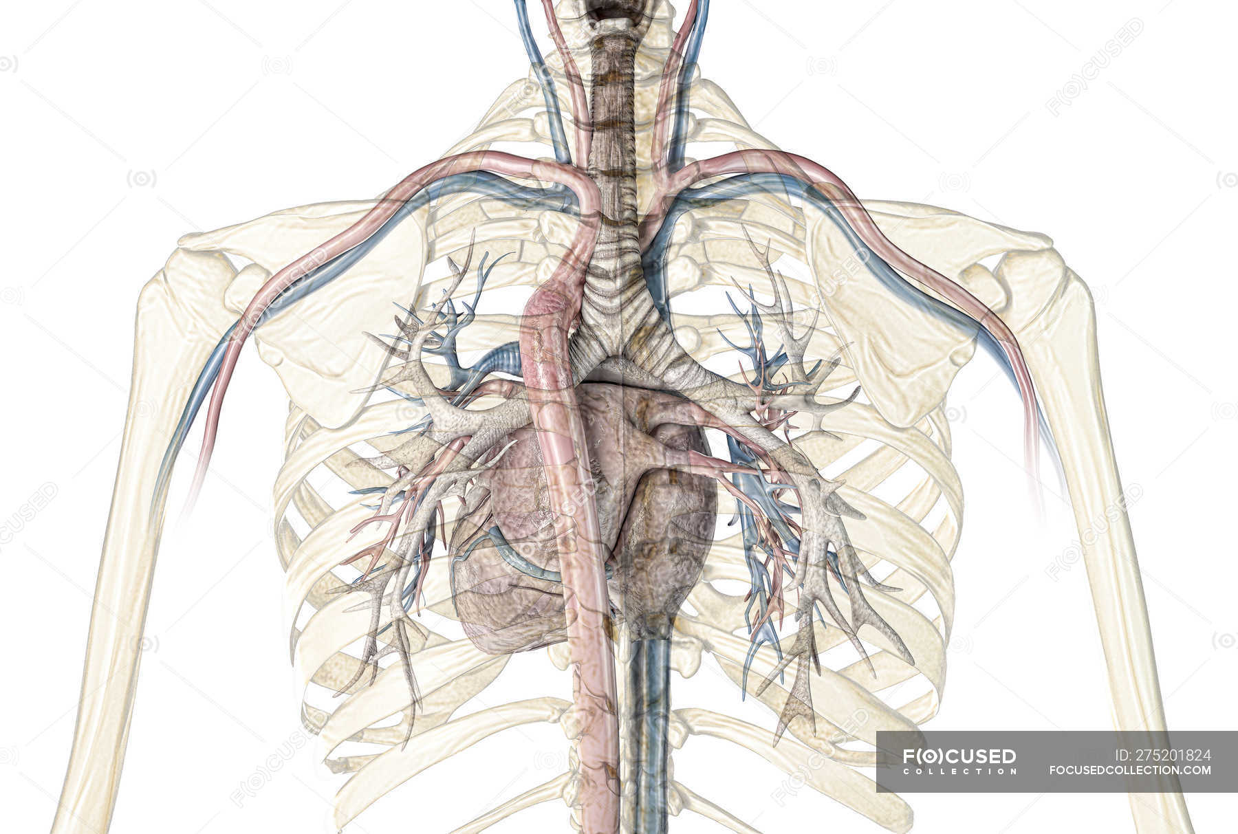
Human Rib Cage Showing Heart With Vessels And Bronchial Tree On White Background Anatomy Tissue Stock Photo 275201824 from st.focusedcollection.com By printing out this quiz and taking it with pen and paper creates for a. Click the image to watch the anatomy of the rib cage video. Includes images, video, and free quiz. Anatomical diagram art print human ribcage butterflys instant digital download a4 printable image pdf jpeg pain+left+side+under+ribs | intro to anatomy 6 learn about anatomy ribs with free interactive flashcards. Pain+left+side+under+ribs | intro to anatomy 6: They articulate with the vertebral column posteriorly, and terminate anteriorly as cartilage (known as costal cartilage). Learn about its function and location as well as conditions that affect the aorta. Human brain functional infographic diagram.
The skull and rib cage.
See more ideas about human anatomy, anatomy, anatomy reference. The distribution of air sacs and the functioning of the avian lung. The rib cage surrounds the lungs and the heart, serving as an important means of bony protection for these vital organs. Learn everything about the ribs with our articles, video tutorials, quizzes, and labeled diagrams there are eleven pairs of external intercostal muscles and these are the most superficial in the area. Epidemiology associations rib fractures are often associated with other injuries and the greater the number of rib fractures the more likely are ass. Rib number 10 is atypical because its head. These are large areas of the cerebral cortex that receive sensory input from multiple. The ribs are a set of twelve paired bones which form the protective 'cage' of the thorax. They articulate with the vertebral column posteriorly, and terminate anteriorly as cartilage (known as costal cartilage). All are attached at the back to the thoracic vertebrae and are numbered from 112 according to the vertebrae they attach to. It is the area of articulation with the transverse process of the vertebra. The first seven are connected behind with the vertebral column and in front. The current morbidity of rib plating is due to the size of the incision required to perform an open procedure.


0 Comments